As we said, there are many types of interstitial lung disease. They have different causes and features, and even different treatments. In some interstitial lung diseases, the interstitium is mostly scarred (aka fibrosed), in others is it mostly inflamed. Sometimes inflammation can lead to fibrosis. Sometimes both fibrosis and inflammation are present.
The goal of your initial visit with the ILD specialist is to determine the best diagnosis that fits with your symptoms and test results. For some, it may take time to get a diagnosis because the ILD may be too early to tell, or it may not fall neatly into any particular category. If this is the case, it is often called "unclassifiable" ILD, and will require periodic monitoring and follow-up with your ILD specialist to determine if it is a progressive disease or not.
There are many ways to categorize interstitial lung diseases. Here is just one:
Some of the most common ILDs we treat and manage in our clinic include:
IPF

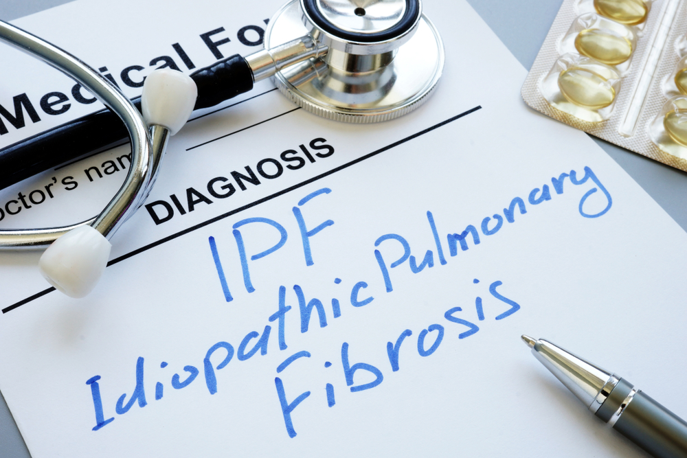
Idiopathic pulmonary fibrosis, or IPF, is a condition that causes progressive scarring of the lungs. "Idiopathic" refers to the fact that the cause of the disease is unknown. (It's important to keep in mind that Idiopathic Pulmonary Fibrosis is just ONE of the many types of pulmonary fibrosis). Fibrous scar tissue builds up in the lungs over time, affecting their ability to provide the body with enough oxygen. The cause of the condition is unknown.
IPF affects more than 100,000 people in the United States, with 30,000 to 40,000 new cases diagnosed each year. Typically the disease is found in people between the ages of 50 and 70 and affects men more frequently than women. Most patients are former smokers. There are no proven risk factors for IPF, but a minority of patients have a family history of lung scarring.
For more information on IPF, please consult the Pulmonary Fibrosis Foundation’s website, a leading resource that provides comprehensive and reliable information on all topics about this disease.
Signs and Symptoms
Symptoms of IPF often appear gradually and include:
- Shortness of breath, particularly during or after physical activity
- Chronic, dry hacking cough
- Crackle sound in the lungs heard through a stethoscope
- Rounding of the fingernails, a condition called clubbing
Symptoms of IPF may mimic those of other diseases that cause lung scarring, so diagnosing IPF often involves ruling out other conditions. Several visits with your doctor may be needed to finalize your diagnosis and treatment approach.
Treatment
Two antifibrotic medications — nintedanib (Ofev) and pirfenidone (Esbriet) — were approved in the fall of 2014 for use in idiopathic pulmonary fibrosis. While these medications are not a cure, they have both been shown to slow the decline of lung function over time. Please see our pharmacologic treatment section for more information on these medications. Please also see our section on antifibrotic therapies under Pharmacologic Treatment.
Ongoing studies of other medications for IPF have shown initial promise, but need more research. For more information on current trials, go here.
Hypersensitivity Pneumonitis
Hypersensitivity pneumonitis (HP) is an interstitial lung disease caused by repeated inhalation of certain fungal, bacterial, animal protein or reactive chemical particles, called antigens. While most people who breathe in these antigens don't develop problems, in some people, the body's immune reaction to these particles causes inflammation of the lung. In some cases, parts of the lungs may become scarred.
It's not known why a minority of people exposed to these antigens develop HP. Their genetics and environment may interact to make them more susceptible to the disease.
HP should not be confused with the more common types of allergies, which are caused by small amounts of proteins in the environment such as dust mites, cat dander, pollen, and grass. Having seasonal or environmental allergies has nothing to do with having or developing HP.
Signs and Symptoms
Hypersensitivity pneumonitis is subdivided into two forms: acute and chronic. Symptoms differ for each form.
Acute Form of HP
The acute form of HP occurs after heavy, often short-term exposure to the antigen. Symptoms appear relatively suddenly and include:
- Fever
- Chills
- Fatigue
- Breathlessness
- Chest tightness
- Cough
If the person is removed from the antigen exposure, the symptoms usually resolve over 24 to 48 hours. Recovery is often complete.
Chronic Form of HP
The chronic form of HP is thought to occur due to longer term, low-level exposure to the antigen, and it often causes more subtle symptoms. Patients with chronic HP often describe chronic symptoms, such as shortness of breath or cough, that have gotten worse. Symptoms may worsen at work, at home or wherever the patient is being exposed to the antigen, but most often, patients with chronic HP haven't had acute episodes. Most patients seen in our clinic have the chronic form of HP.
Diagnosis
In addition to history, physical examination, and the various tests that might be ordered to help with diagnosis, a thorough review of potential occupational and environmental exposures to antigens as well as a detailed home and work history are particularly essential when diagnosing HP. This will include exposures to mold, birds and bird products, such as down. Often times, you may be given a home checklist (LINK) to fill out and send back to us, asking you to thoroughly evaluate your home for any potential exposures.
For some, a convincing exposure or antigen might never be discovered. While frustrating, this is not unusual for nearly half of patients who are diagnosed with HP.
Treatment
Treating hypersensitivity pneumonitis (HP) involves both identifying and removing the antigen that's causing the condition, and taking anti-inflammatory medication.
Removing the Antigen
If the inhaled antigen can be recognized and removed, the lung inflammation in acute HP is often reversible. If you have chronic HP, however, the inflammation may persist even when the antigen is removed. If the antigen can't be identified, you may need to change your work or home environment, if possible.
Medication Therapy
If you don't improve or continue to worsen, we may recommend anti-inflammatory medications. Prednisone is the mainstay of medication therapy and is often very effective. If you require long-term medication or don't tolerate prednisone, you may need to take an alternative medication, such as mycophenolate or cyclophosphamide.
Connective Tissue Disease - ILD
Connective tissue disease associated with interstitial lung disease, or CTD-ILD, is a lung condition that affects a small number of patients with connective tissue disease. Examples of connective tissue diseases — also known as rheumatologic, collagen vascular or autoimmune diseases — include scleroderma, rheumatoid arthritis, Sjogren's syndrome, systemic lupus erythematosus, polymyositis, dermatomyositis and mixed connective tissue disease.
Patients are often diagnosed with the connective tissue disease first and develop CTD-ILD later, although in some cases, the opposite occurs. For those in whom interstitial lung disease is the first manifestation of connective tissue disease. If this is the case, we may refer you to a rheumatologist for further evaluation.
CTD-ILD causes inflammation or scarring (fibrosis) of the lungs. The exact cause of lung damage is unknown.
Signs and Symptoms
Some patients with CTD-ILD don't have symptoms. For others, common symptoms include:
- Shortness of breath with activity
- Cough
- Fatigue
- "Crackle" sound heard when listening to the chest with a stethoscope
- Symptoms of a connective tissue disease, such as joint pain and swelling, rash, dry eyes, dry mouth and acid reflux
Treatment
CTD-ILD is treated with anti-inflammatory or immunosuppressive medications. You may recognize some or all of these medications if they were prescribed to you for your connective tissue disease.
The most common medications used to treat CTD-ILD are immunosuppressive medications like steroids and/or steroid sparing agents. For certain CTD-ILD diagnoses such as scleroderma, antifibrotic medications may be indicated. Please see our pharmacologic treatment session for more information on these medications
Sarcoidosis
Sarcoidosis is a disorder that causes inflamed tissue, called nodules or granulomas, to develop in the body's organs, most often the lungs. It can also affect the skin, eyes, nose, muscles, heart, liver, spleen, bowel, kidney, testes, nerves, lymph nodes and brain. Nodules in the lungs can lead to narrowing of the airways and inflammation, also called fibrosis, of lung tissue.
Sarcoidosis affects people of all ages, races, and gender, though it most commonly occurs in people between 20 to 40 years old. Children are rarely diagnosed with the disease. In very few cases, more than one family member is affected. African-Americans are three to four times more likely to have sarcoidosis and may have a more severe form of the disease than people of European descent.
Signs and Symptoms
Symptoms of sarcoidosis may vary from person to person, and depend on the organs affected. Frequently, the condition causes mild symptoms and resolves on its own without treatment. In approximately half of all patients, sarcoidosis is detected on a routine chest X-ray before any symptoms develop. The most common symptoms of sarcoidosis involving the lungs include:
- Cough
- Shortness of breath
- Chest pain, which is usually a vague tightness of the chest, but can occasionally be severe and similar to the pain of a heart attack
- Fatigue
- Weakness
- Fever
- Weight loss
Treatment
The cause of sarcoidosis is unknown at this time. Therefore, there is no specific treatment to cure the condition. Fortunately, in many cases, sarcoidosis does not require treatment because the nodules seen on your CT scan gradually resolve on their own and leave behind few, if any, signs of inflammation or other complications.
However, treatment is necessary in some cases. Medications are available that effectively suppress symptoms and help reduce lung inflammation, the impact of nodules and prevent the development of lung fibrosis.
Corticosteroids, usually prednisone, are particularly effective in reducing inflammation and are typically the first drugs used in the treatment of sarcoidosis. In patients with mild symptoms, such as skin lesions, eye inflammation, or cough, topical steroid therapy with creams, eye-drops or inhalers may be sufficient to control the disease. When necessary, oral steroids are generally prescribed for six to twelve months. In most cases, a relatively high dose is prescribed at first, followed by a slow taper to the lowest effective dose.
Symptoms, especially cough and shortness of breath, generally improve with steroid therapy. Relapses may occur after treatment with steroids has ended, but typically respond to repeated steroid treatment. Patients who improve and remain stable for more than a year after the end of treatment have a low rate of relapse.
Researchers continue to examine the role of steroids in the treatment of sarcoidosis, with some addressing the question of what effect they may have on the long-term course of the disease. However, in general, steroid therapy remains the leading treatment for sarcoidosis.
Alternative medications are used in patients who cannot tolerate steroids, do not respond to steroids or wish to lower the dose of steroids. These are referred to as steroid sparing agents, and more information can be found here. There are some medications that are commonly used in sarcoidosis that are unique from other interstitial lung diseases. These include:
- Methotrexate – This medication reduces inflammation and suppresses the immune system and is commonly used in the treatment of autoimmune disease or certain cancers.
- Cyclophosphamide (Cytoxan) – Though less commonly used these days, this medication can be used in conjunction with steroids in patients whose condition is worsening despite treatment.
- Hydroxychloroquine (Plaquenil) – These medications, known as immunosuppressive and anti-parasitic medications, are used to treat sarcoidosis of the skin and lungs.
- Colchicine – This medication is most commonly used to treat gout and is sometimes prescribed to treat sarcoidosis-related arthritis for its anti-inflammatory effects.
A number of other medications are currently being investigated for the treatment of sarcoidosis. For more information about ongoing clinical trials in sarcoidosis, please refer to this page.
