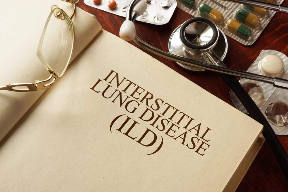
Interstitial lung disease (ILD) is a general term for a group of conditions that cause scarring, called fibrosis, or inflammation of the lungs and gradually interferes with breathing. The diagnosis and management of ILD is often complex and requires input from doctors across several areas of expertise. The UCSF Interstitial Lung Disease Program evaluates all forms of interstitial lung disease. See our Different Types of ILD section for more information.
In order to understand what interstitial lung disease (ILD) is and how it affects your health, it is important to understand our need for oxygen and how the lungs work.
Every organ and tissue and cell in our bodies needs oxygen to work right and to be healthy. Without enough oxygen, your body functions poorly and you can feel symptoms such as fatigue, poor exercise tolerance or shortness of breath. How do our organs, tissues and cells get oxygen? It is the job of the lungs to take oxygen molecules from the air and deliver them to the bloodstream, where they are then carried to every part of the body.
Here's how that happens. When you take a breath, air first travels into the lungs via the airways, which begin with the trachea, then divide into two mainstem bronchi, which in turn divide into smaller airways, which branch and branch and branch into smaller and smaller airways. This branching occurs 30-35 times, resulting in a vast network of microscopic airways throughout the lungs.

Diseases of these airways are ones you have probably heard of, such as asthma, emphysema, COPD, and bronchitis. These are problems of the flow of air in and out of the lungs; they are called obstructive airways diseases because the flow of air is obstructed. Most people have heard of these airways diseases because they are more common than interstitial lung diseases. Airways diseases are often treated with inhalers such as albuterol and steroids, which open up the airways so that air flows better.

But let’s look at some other structures in the lungs to find out what interstitial lung disease is. At the ends of all those microscopic airways are tiny airsacs – also called alveoli. Our lungs have hundreds of millions of these airsacs. In fact, if you look at a slice of lung tissue under a microscope, it looks like a very fine sponge – you’re looking at the all the airsacs.
What happens in these airsacs? This is where oxygen molecules that we breathe in from the outside air goes into the bloodstream – remember, this is the whole purpose of breathing.
So, we breathe in air, it goes down the airways, into the airsacs, then oxygen molecules from that air cross into the bloodstream, where red blood cells with hemoglobin inside of them float by, grab the oxygen molecules, and carry them off to all our organs, tissues and cells.

But the oxygen molecules have to cross a tissue between the airsac and the blood vessel called the interstitium before they go into the bloodstream. The interstitium is a highly specialized tissue made up of epithelial cells from the lining if the airsac, endothelial cells from the lining of the blood vessel, and something called matrix in between. The interstitium facilitates what we call gas exchange in the lungs – oxygen in, carbon dioxide out.
When the interstitial tissue is injured or altered in some way, it’s hard for oxygen molecules to cross into the bloodstream. When this happens, it’s called an interstitial lung disease. Other parts of the lung may also be affected when you have an ILD, including the airways and blood vessels.
There are about 200 (or many, many) reasons people develop interstitial lung disease. It is the task of the interstitial lung disease team to figure out the specific cause of inflammation and/or scarring in the interstitium, and how best to treat it.
Note: It is possible to have both an airways disease, such as COPD or asthma, and an interstitial lung disease.